The Human Eye
NB. : The eye : an analog-digital converter ...
When light get to cones and rods cells, it creates a electric potential. Above
a given level (sensitivity of the cell), a action potential is transmitted from
one nervous cell to another. Height and length of the action potential are constant,
only the frequency (number of potential) varies regarding the intensity of the
the stimulus (here the light).
At that level, we already may notice that two things may inform the human of the distance of an object : the contraction of ciliary muscles (focus), and the orbital muscle that position the orientation of the eye (stereoscopic view).
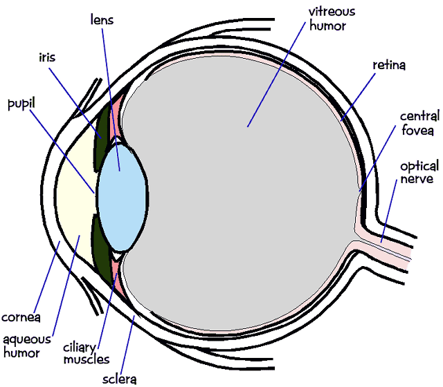
The eye is essentially an opaque eyeball filled with a water-like fluid. In the front of the eyeball is a transparent opening known as the cornea. The cornea is a thin membrane which has an index of refraction of approximately 1.38. The cornea has the dual purpose of protecting the eye and refracting light as it enters the eye. After light passes through the cornea, a portion of it passes through an opening known as the pupil. Rather than being an actual part of the eye's anatomy, the pupil is merely an opening. The pupil is the black portion in the middle of the eyeball. It's black appearance is attributed to the fact that the light which the pupil allows to enter the eye is absorbed on the retina (and elsewhere) and does not exit the eye. Thus, as you sight at another person's pupil opening, no light is exiting their pupil and coming to your eye; subsequently, the pupil appears black.
Like the aperture of a camera, the size of the pupil opening
can be adjusted by the dilation of the iris. The iris is the
colored part of the eye - being blue for some people and brown for others (and
so forth); it is a diaphragm which is capable of stretching and reducing the
size of the opening. In bright-light situations, the iris is dilated to reduce
the size of the pupil and limit the amount of light which enters the eye; and
in dim-light situations, the iris adjusts its size so as to maximize the size
of the pupil and increase the amount of light which enters the eye.
Light which passes through the pupil opening, will enter the crystalline lens. The crystalline lens is made of a fibrous, jelly-like material which has an index of refraction of 1.44. Unlike the lens on a camera, the lens of the eye is able to change its shape and thus serves to fine-tune the vision process. The lens is attached to the ciliary muscles. These muscles relax and contract in order to change the shape of the lens. By carefully adjusting the lenses shape, the ciliary muscles assist the eye in the critical task of producing an image on the back of the eyeball.
The inner surface of the eye is known as the retina. The retina contains the rods and cones which serve the task of detecting the intensity and the frequency of the incoming light. An adult eye is typically equipped with 120 million rods which detect the intensity of light and 6 million cones which detect the frequency of light. These rods and cones send nerve impulses to the brain. The nerve impulses travel through a network of nerve cells; there are as many as one-million neural pathways from the rods and cones to the brain. This network of nerve cells is bundled together to form the optic nerve on the very back of the eyeball.
Each part of the eye plays a distinct part in enabling humans to see. The ultimate goal of such an anatomy is to allow humans to focus images on the back of the retina.
Extract from http://www.physicsclassroom.com/Class/refrn/U14L6a.html